A. Major Structures involved in Hearing:
 |
|
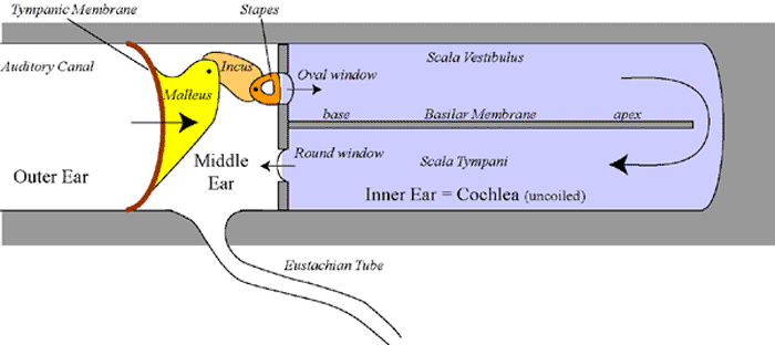
1. Outer Ear: Consists of the pinna (= auricle) and the external auditory canal. The pinna is important for the collection (= amplifies the sound signal) and to determine the direction (more sensitive to signals from the front then from the back of the head) of the sound waves. Some animals can even rotate their ears towards the source of the sound (dogs, horses, camels). The auditory canal is important for the conduction of sound waves from outside to the tympanic membrane. If the canal is obstructed (wax, foreign bodies) then less sound waves will reach the ears leading to deafness. |
2. Middle Ear: Is an air-filled space between outer ear and inner ear. Inside, there are three ossicles; the malleus, the incus and the stapes:
These ossicles work as levers; thereby magnifying the amplitude of the sound waves (approximately 20x). |
3. The Eustachian Tube: Connects the middle ear to the pharynx (and therefore to the outside world). This communication is necessary to keep the pressure inside the middle ear equal to that outside the Tympanic membrane. Otherwise (if there is a pressure difference), this will cause the tympanic membrane to be pushed either in or out of the middle ear. This imbalance will make a person deaf and can be very painfull. The tympanic membrane works best when the pressure at both sides is equal. Well known is the problem in airplanes, especially during take off or during landing (crying children). |
4. The Attenuation Reflex: The ossicles are very delicate and, when there is a loud noise, they need protection. Two muscles are attached to the ossicles: 1. the stapedius muscle 2. the tensor tympani. These contract by reflex, when there is a loud noise, to pull at the ossicles and to prevent them of getting dislodged. |
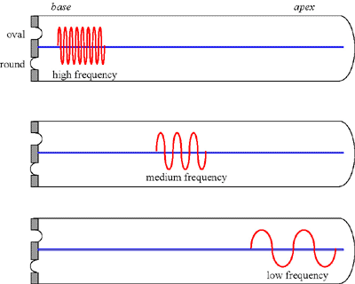
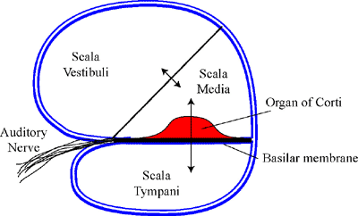
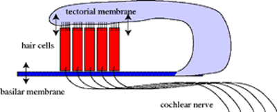
5. The Cochlea: The cochlea is a spiral shaped structure with a base, close to the middle ear, and an apex, at the distal end. In the diagram above, the cochlea is unrolled (= uncoiled) as a straight tube. The inside of the cochlea is subdivided into three scala's: the scala vestibuli, the scala media and the scala tympani. The scala media and vestibuli work together. Between these two and the scala tympani, there is the basilar membrane. On this basilar membrane, the sensor of the ear, the organ of Corti is located. |
6. Sound Transduction: Sound waves propagate through the outer ear and vibrate the Tympanic membrane. The membrane vibrations make the ossicles vibrate (and increase the amplitude). Finally, the stapes vibrates against the membrane of the oval window, which will make the fluid in the vestibular scala vibrate. These vibrations will propagate to the apex and then down along the tympanic scala. Therefore the basilar membrane will first move according to the upper pressure and, a little bit later, according to the lower pressure vibrations. |